Overview
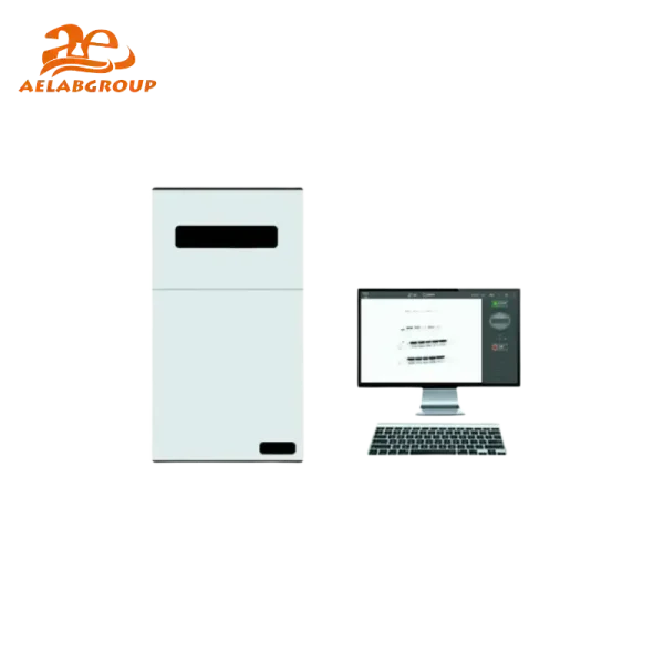
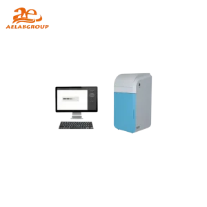
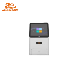
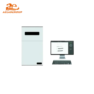
AELAB Automatic Gel Imaging and Analysis System is a highly integrated and fully automated gel imaging system. The system interface is simple and practical, and the main interface has shooting button and time control to guide the user. It is very easy to operate, no need to have manual and tedious debug, it is your best choice.
The AELAB AE-GEL100/200 system delivers high-resolution CCD imaging, high sensitivity, automatic focus, and high-performance optical filtering for nucleic acid dye detection. With UV, white light, and optional Epi-UV illumination, the system supports precise visualization across common fluorescence assays.
⚙️ Design & Construction
The AE-GEL100 and AE-GEL200 incorporate high-QE CCD sensors, providing enhanced photon detection for weak fluorescence signals. A motorized 8–48 mm F1.2 lens ensures precise focus control, while multilayer coating filters improve signal-to-noise performance. The system integrates UV (302 nm), Epi-UV (254/302/365 nm optional), white-light LED illumination, and a UV-to-white sample plate for SDS-PAGE gel imaging. A durable enclosure protects samples from ambient light and optimizes imaging uniformity.
🔬 Working Principle / Testing Method
The instrument captures fluorescence or white-light images by directing UV or LED illumination onto DNA, RNA, or protein gels. The CCD sensor converts emitted fluorescence into digital signals with 16-bit depth (0–65535 levels), enabling quantitative analysis of gel bands. Exposure adjustment from milliseconds to extended durations allows imaging of low-intensity targets. Binning modes (1×1, 2×2, 4×4 for AE-GEL200) further enhance sensitivity during low-light conditions.
💡 Key Advantages
- High-efficiency QE sensor for low-signal nucleic acid fluorescence imaging
- Motorized autofocus for rapid and reproducible image acquisition
- Multi-wavelength UV/Epi-UV options supporting diverse fluorescent dyes
- High dynamic range 16-bit imaging suitable for quantitative analysis
- Integrated UV-to-white plate enabling SDS-PAGE and protein visualization
- Advanced filter options for EB, SYBR Safe, SYBR Green, GelRed, and other dyes
- Simple user interface enabling fast imaging without manual calibration
🔗 Connectivity & Integration
The system supports PC connectivity for real-time display, image capture, and data export. Compatible analysis software enables band quantification, molecular weight estimation, and documentation. USB data transfer facilitates GLP-compliant archiving and laboratory report generation.
🧠 Model Insight
| Model |
Specifications |
| AE-GEL100 |
2592×1944 CCD, >65% QE, 1ms–3000ms exposure, 1×1 binning, 16-bit depth, UV/white light imaging |
| AE-GEL200 |
3000×4000 CCD, >75% QE, 1ms–120min exposure, 1×1/2×2/4×4 binning, 16-bit depth, UV/white light imaging |
🧪 Testing Applications
- DNA gel imaging (EB, SYBR Green, SYBR Safe, GelRed, Fluorescein dyes)
- RNA visualization and quantitative fluorescence documentation
- SDS-PAGE protein analysis and band documentation
- Thin-layer chromatography imaging
- Colony counting under white-light illumination
- Nucleic acid quantification workflows in molecular biology laboratories
Compliance & Quality Assurance
The AE-GEL Series complies with international laboratory quality standards, ensuring reproducible imaging for GLP environments. Optical performance, UV illumination stability, and electronic components undergo ISO-aligned manufacturing and verification procedures.