AELAB | Laboratory Equipment | Microtome
Microtome
Introduction
A Microtome is a precision instrument essential for creating thin tissue sections for microscopic analysis. Commonly used in histology, pathology, and biological research, microtomes ensure consistent section thickness, enabling accurate observation at the cellular and structural levels for diagnostics and advanced scientific studies.
What Is Microtome?
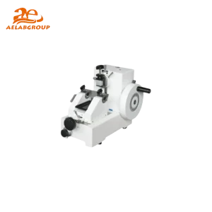
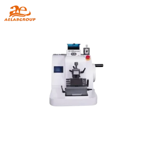
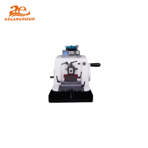
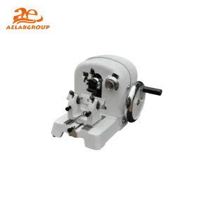
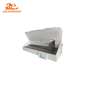
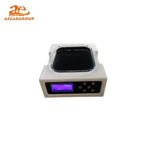
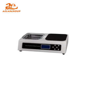
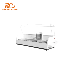
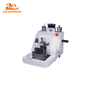
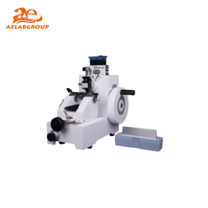
A microtome is a laboratory device designed to slice samples into ultra-thin sections for microscopic examination. It consists of a sharp blade, specimen holder, and an advancing mechanism that moves the sample toward the blade with controlled precision. Microtomes are indispensable in histopathology, forensic science, and materials research, where uniform section thickness ensures reproducible analysis and imaging quality.
Devices in This Category
Rotary Microtome
Cryostat Microtome
Ultramicrotome
Vibrating Microtome
Sliding Microtome
Handheld Microtome
Technical Features and Specifications
FeatureDetails
Section Thickness Range0.1 – 60 µm (depending on model)
Cutting ModeManual, semi-automatic, or fully automatic
Blade TypeDisposable steel, reusable glass, or diamond
Specimen RetractionAutomatic retraction to reduce wear on the blade
Specimen SizeUp to 50×60 mm (standard tissue block)
ErgonomicsAdjustable handwheel, anti-fatigue design, easy cleaning
Precision ControlMicrometer-driven or digital feed adjustment
Safety FeaturesBlade guards, locking mechanisms, emergency stop
Benefits
Produces thin, consistent sections ideal for microscopy.
Supports a wide range of biological and material samples.
Enhances diagnostic precision in histopathology.
Reduces processing time with semi- and fully automated systems.
Improves reproducibility and research quality.
Applications and Tests
🔬 Molecular Biology
Preparation of tissue sections for in situ hybridization
Sample slicing for gene expression and localization studies
Sectioning embedded tissues for microscopic imaging
🧪 Clinical Diagnostics
Biopsy sectioning for cancer diagnosis and histopathology
Frozen section preparation during surgical procedures
Embedding and sectioning for immunohistochemistry (IHC)
🏭 Industrial & Food Testing
Sectioning polymer or composite materials for structural analysis
Microscopic inspection of coatings and laminates
Quality assurance of packaging and film layers
🌱 Environmental & Agricultural Labs
Plant tissue sectioning for cellular and anatomical studies
Soil and seed cross-section preparation for microscopy
Biomaterial sample slicing for ecological research
Microtome vs. Cryostat vs. Ultramicrotome
FeatureMicrotomeCryostatUltramicrotome
Sample StateParaffin-embeddedFrozenResin-embedded
Section Thickness1–60 µm5–30 µm50–100 nm
Use CaseRoutine pathologySurgical diagnosticsElectron microscopy
Automation LevelManual to automaticTemperature-controlled manualUltrahigh precision motorized
CostModerateHighVery High
Expert Tips for Choosing and Using Microtomes
Select the correct microtome type based on sample state—frozen, paraffin, or resin-embedded.
Calibrate thickness settings before every use to ensure uniform sectioning.
Use appropriate disposable or diamond blades for your specimen hardness.
Employ anti-roll plates or section stretchers to achieve smooth ribbons.
Clean and lubricate mechanical components routinely for precision and safety.
Maintenance Best Practices
Clean the blade holder and stage after each use with lint-free tissue.
Check and sharpen or replace blades regularly for consistent performance.
Inspect moving parts and lubricate the handwheel mechanism periodically.
Keep specimen clamps and anti-roll devices free of paraffin or debris.
Follow manufacturer calibration and servicing intervals for optimal longevity.
FAQ
Q: What is a microtome used for?
A: A microtome is used to cut extremely thin tissue or material sections for microscopic examination, essential in pathology and research labs.
Q: How thin can a microtome cut?
A: Depending on the type, microtomes can produce sections from 0.1 µm to 60 µm thick, while ultramicrotomes can reach 50–100 nm for electron microscopy.
Q: What is the difference between a microtome and a cryostat?
A: A microtome cuts paraffin-embedded samples at room temperature, while a cryostat performs similar sectioning on frozen tissues within a cooled chamber.
Q: How do I maintain my microtome?
A: Regular cleaning, blade replacement, lubrication, and calibration are essential to ensure long-term accuracy and operator safety.